Shoulder Labral Tear Mri, Artifacts And Pitfalls In Shoulder Magnetic Resonance Imaging
Shoulder labral tear mri Indeed lately has been hunted by users around us, maybe one of you. People are now accustomed to using the net in gadgets to view image and video data for inspiration, and according to the title of the post I will talk about about Shoulder Labral Tear Mri.
- The Radiology Assistant Shoulder Instability Mri
- Slap Tear Symptoms Diagnosis And Treatment Everything You Need To Know Dr Nabil Ebraheim Youtube
- Shoulder Labral Tears Mri
- Diagnosis Glenoid Labral Tear Pogo Physio Gold Coast
- Https Encrypted Tbn0 Gstatic Com Images Q Tbn 3aand9gctn84pmstuoo5bd9ifoy8dm 7wrgizvgi6g6pibky2vceebygtv Usqp Cau
- Shoulder Labral Tears Mri
Find, Read, And Discover Shoulder Labral Tear Mri, Such Us:
- Diagnosis Glenoid Labral Tear Pogo Physio Gold Coast
- Glenohumeral Instability Radsource
- My Shoulder Superior Labrum Is Torn Do I Need Surgery Shoulder Elbow
- What Can The Radiologist Do To Help The Surgeon Manage Shoulder Instability
- Shoulder Labral Tears Mri
If you re searching for Jungkook Birthday Cake 2020 you've reached the perfect place. We have 104 images about jungkook birthday cake 2020 including images, pictures, photos, wallpapers, and much more. In such page, we also have variety of images out there. Such as png, jpg, animated gifs, pic art, logo, blackandwhite, translucent, etc.
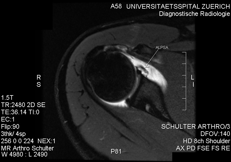
Anteriorinferior labral tear with stripping of the periosteum compatible with a soft bankart and a three part lesion.

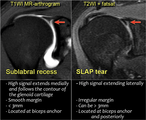
Jungkook birthday cake 2020. When an mri with contrast is ordered contrast is injected into the vein while the arthrogram injects contrast directly into the joint under fluoroscopy guidance. This means that mr arthrography with the arm in the neutral position may fail to detect the labral tear. Coronal diagram of the superior labrum depicts three entities that may produce linear hyperintensities that mimic a superior labral tear.
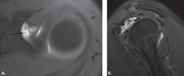
Labral injury extends anteriorly from approximately the 10 oclock position to the 6 oclock position. The mri allows accurate assessment of any pathologic changes of the structures of the shoulder including the glenoid labrum the humeral head the articular cartilage and the rotator cuff. Glenoid labral tears are the injuries of the glenoid labrum and a possible cause of the shoulder pain.
Rotator cuff tears the aber view is also very useful for both partial and full thickness tears of the rotator cuff. Cartilage undercutting c a pseudoslap lesion between the lhbt and superior labrum p and a sublabral recess or sulcus r. Clinical presentation patients with labral tears may present with a wide range of symptoms depends on the injury type which are often non.
Slap tears involve the superior glenoid labrum where the long head of biceps tendon inserts. S common configuration of an actual slap lesion. On images of the shoulder with the arm in a neutral position the torn labrum may be held in its normal anatomic position by the intact scapular periosteum which thereby prevents contrast media from entering the tear.
T2 star gradient recall echo images are employed in the assessment of the labrum and for detection of substances that produce susceptibility effects such as calcium hydroxyapatite or loose surgical hardware. In addition chronic tiny superior labral injury. Unlike bankart lesions and alpsa lesions they are uncommonly 20 associated with shoulder instability 5.
The purpose of this article is to review this subject to describe problems related to normal anatomy and variants of the superior and anterosuperior portions of the labrum to perform a critical analysis of the current 10 grade slap lesion classification and mechanisms of injury from the perspective of mri and to describe an mri approach to the diagnosis of such lesions. In the acute setting they are most frequently seen in. Mri shoulder protocols typically involve fat saturated proton density images that are sensitive to internal derangement.
Rule out labral tear to rule out a labral tear an mri arthrogram needs to be ordered not an mri with contrast. They can extend into the tendon involve the glenohumeral ligaments or extend into other quadrants of the labrum.
More From Jungkook Birthday Cake 2020
- Bts Billboard 2019
- Yoongi Mirror Selfie 2020
- Jingle Bells Clipart
- Jimin Bts Gif Hd
- Bts Grammys 2020 Vlive
Incoming Search Terms:
- Alpsa Lesion Wikipedia Bts Grammys 2020 Vlive,
- Orthopedic Center Of St Louis Bts Grammys 2020 Vlive,
- The Radiology Assistant Shoulder Mr Instability Bts Grammys 2020 Vlive,
- Role Of Conventional Mri And Mr Arthrography In Evaluating Shoulder Joint Capsulolabral Ligamentous Injuries In Athletic Versus Non Athletic Population Sciencedirect Bts Grammys 2020 Vlive,
- The Shoulder Radiology Key Bts Grammys 2020 Vlive,
- Welcome To Ecronicon Bts Grammys 2020 Vlive,


