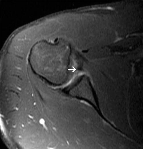
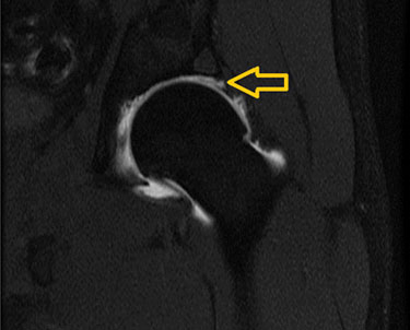
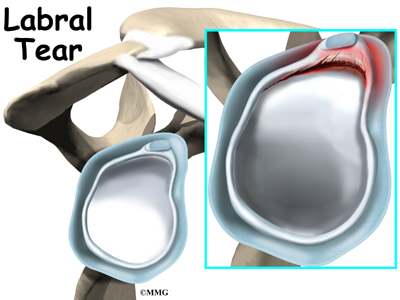
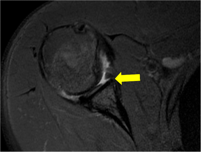
Shoulder Labral Tear Mri Images, Displaced Fragment Of A Bucket Handle Tear Of Glenoid Labrum Seen At Download Scientific Diagram
Shoulder labral tear mri images Indeed recently has been sought by users around us, maybe one of you personally. People now are accustomed to using the net in gadgets to see image and video information for inspiration, and according to the title of the article I will discuss about Shoulder Labral Tear Mri Images.
- Posterior Shoulder Instability With Labral Tear International Congress For Joint Reconstruction
- Magnetic Resonance Imaging Evaluation Of Meniscoid Superior Labrum Normal Variant Or Superior Labral Tear
- Posterior Labral Tear Shoulder Elbow Orthobullets
- The Radiology Assistant Shoulder Mr Instability
- Displaced Fragment Of A Bucket Handle Tear Of Glenoid Labrum Seen At Download Scientific Diagram
- Slap Tear Type Iii Radiology Case Radiopaedia Org
Find, Read, And Discover Shoulder Labral Tear Mri Images, Such Us:
- Arthroscopic Repair Of Posterior Labral Tear With Paralabral Cyst Decompression
- The Radiology Assistant Shoulder Instability Mri
- Posterior Labral Tear Shoulder Elbow Orthobullets
- Slap Tears And Biceps Tendon Injuries Huang Orthopaedics
- The Epidemiology Of Mri Detected Shoulder Injuries In Athletes Participating In The Rio De Janeiro 2016 Summer Olympics Bmc Musculoskeletal Disorders Full Text
If you re searching for Bts Spring Day Lyrics Romanized you've reached the ideal location. We have 104 images about bts spring day lyrics romanized including pictures, photos, photographs, backgrounds, and more. In these page, we additionally have variety of images out there. Such as png, jpg, animated gifs, pic art, symbol, black and white, transparent, etc.
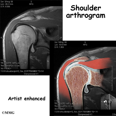
Allowing intra articular contrast to get between the labral tear and the glenoid.

Bts spring day lyrics romanized. The role of diagnostic imaging in the evaluation of shoulder pain is to guide clinical management. T2 star gradient recall echo images are employed in the assessment of the labrum and for detection of substances that produce susceptibility effects such as calcium hydroxyapatite or loose surgical hardware. However there are several mr imaging characteristics that can aid the radiologist in diagnosis.
The glenoid labrum an important static stabilizer of the shoulder joint has several normal labral variants that can be difficult to discriminate from labral tears and is subject to specific pathologic lesions anteroinferior posteroinferior and superior labral anteroposterior lesions with characteristic imaging features. Mri shoulder protocols typically involve fat saturated proton density images that are sensitive to internal derangement. Mri is helpful in differentiating this syndrome from an actual tear.
Unlike bankart lesions and alpsa lesions they are uncommonly 20 associated with shoulder instability 5. Inflammation and swelling then follows and and leads to weakness decreased range of motion and pain in the shoulder area. Slap tears involve the superior glenoid labrum where the long head of biceps tendon inserts.
Imaging in three planes is advisable and additional orthogonal planes may be included in the protocol for a detailed assessment of the lesion. Mri gallery shoulder mri musculoskeletal imaging mri of the shoulder. On conventional mr labral tears are best seen on fat saturated fluid sensitive sequences.
Mri evaluation of the shoulder tendon allows for the assessment of the tendons surrounding the shoulder known as the rotator cuff as well as assess for trauma to the cartilage and labrum the latter in cases of episodes of instability such as following dislocation. In the presence of a rotator cuff tear imaging can determine whether the tear is full thickness or partial thickness and thus help the clinician decide between operative or nonoperative treatment if surgical treatment is decided imaging can be used further to plan the surgical. They can extend into the tendon involve the glenohumeral ligaments or extend into other quadrants of the labrum.
Shoulder anatomy mri normal anatomy variants and checklist. Scroll through the images and notice the unattached labrum at the 12 3 oclock position at the site of the sublabral foramen. The process of diagnosing a labral tear is complicated by the various pitfalls in imaging diagnosis often present in the superior labrum 1734 which can make this a challenging task for the radiologist.
In the acute setting they are most frequently seen in. On images of the shoulder with the arm in a neutral position the torn labrum may be held in its normal anatomic position by the intact scapular periosteum which thereby prevents contrast media from entering the tear.
More From Bts Spring Day Lyrics Romanized
- Bts Jimin Tattoo Hand
- Jungkook Cute Wallpaper Bts
- Bts Tattoo Ideas Printable
- Taehyung Dynamite Haircut
- Bts Dynamite Lyrics Quotes
Incoming Search Terms:
- Mr Arthrogram For Shoulder Microinstability And Hidden Lesions Sciencedirect Bts Dynamite Lyrics Quotes,
- Charter Mr Arthrogram Torn Shoulder Labrum 3 Charter Radiology Bts Dynamite Lyrics Quotes,
- The Radiology Assistant Shoulder Instability Mri Bts Dynamite Lyrics Quotes,
- Slap Tear Symptoms Diagnosis And Treatment Everything You Need To Know Dr Nabil Ebraheim Youtube Bts Dynamite Lyrics Quotes,
- Magnetic Resonance Imaging Evaluation Of Meniscoid Superior Labrum Normal Variant Or Superior Labral Tear Bts Dynamite Lyrics Quotes,
- Https Www Ajronline Org Doi Pdf 10 2214 Ajr 08 1097 Bts Dynamite Lyrics Quotes,

