Right Shoulder Labral Tear Mri, The Radiology Assistant Shoulder Instability Mri
Right shoulder labral tear mri Indeed lately is being sought by users around us, perhaps one of you. People now are accustomed to using the internet in gadgets to see video and image information for inspiration, and according to the title of the post I will discuss about Right Shoulder Labral Tear Mri.
- Role Of Conventional Mri And Mr Arthrography In Evaluating Shoulder Joint Capsulolabral Ligamentous Injuries In Athletic Versus Non Athletic Population Sciencedirect
- Mr Arthrography And Ct Arthrography In Sports Related Glenolabral Injuries A Matched Descriptive Illustration Springerlink
- Bankart Lesion M24 119 718 31 Eorif
- Shoulder Labral Tear What You Should Know And What Surgeons Won T Say
- The Radiology Assistant Shoulder Anatomy Mri
- Mr Arthrogram Of The Right Shoulder Shows Irregular Tear Of The Download Scientific Diagram
Find, Read, And Discover Right Shoulder Labral Tear Mri, Such Us:
- Slap Tears And Biceps Tendon Injuries Huang Orthopaedics
- Glenohumeral Instability Radsource
- Arthroscopic Repair Of A Suspected Type Ii Slap Tear International Congress For Joint Reconstruction
- The Radiology Assistant Shoulder Anatomy Mri
- Shoulder Labral Tears Mri
If you are looking for Bts Stay Gold Wallpaper For Laptop you've reached the ideal location. We ve got 104 graphics about bts stay gold wallpaper for laptop including images, photos, photographs, backgrounds, and much more. In such page, we also have number of graphics available. Such as png, jpg, animated gifs, pic art, symbol, blackandwhite, transparent, etc.
To rule out a labral tear an mri arthrogram needs to be ordered not an mri with contrast.

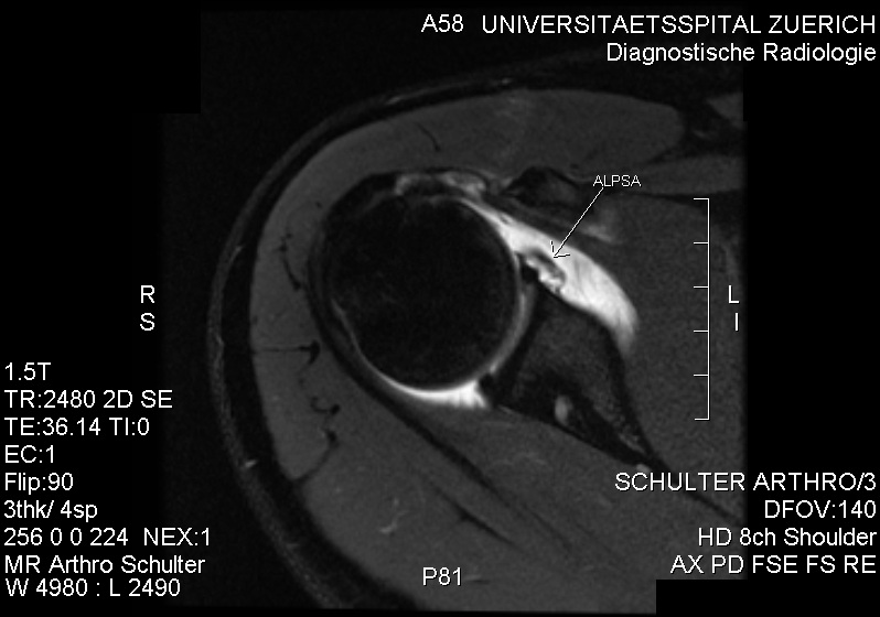
Bts stay gold wallpaper for laptop. Rule out labral tear. The abduction external rotation aber view is excellent for assessing the anteroinferior labrum at the 3 6 oclock position where most labral tears are located. Mri shoulder protocols typically involve fat saturated proton density images that are sensitive to internal derangement.
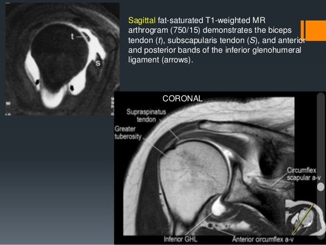
It is composed of two articulations. In the aber position the inferior glenohumeral ligament is stretched resulting in tension on the anteroinferior labrum allowing intra articular contrast to get between the labral tear and the glenoid. Coronal t1 weighted fat suppressed image 60015 obtained posterior to level of a reveals labral tear arrow characterized as slap ii tear.
In the acute setting they are most frequently seen in. They can extend into the tendon involve the glenohumeral ligaments or extend into other quadrants of the labrum. This means that mr arthrography with the arm in the neutral position may fail to detect the labral tear.
The shoulder joint is a joint that connects the upper limb to the axial skeleton. Unlike bankart lesions and alpsa lesions they are uncommonly 20 associated with shoulder instability 5. On images of the shoulder with the arm in a neutral position the torn labrum may be held in its normal anatomic position by the intact scapular periosteum which thereby prevents contrast media from entering the tear.
When an mri with contrast is ordered contrast is injected into the vein while the arthrogram injects contrast directly into the joint under fluoroscopy guidance. Hh humeral head g glenoid. Has to determine if there is a labrum tear in only one to two and at most three slices.
Discussion our study is the first to evaluate superior labra with noncontrast shoulder mris in 45 to 60 year old asymptomatic people. This means that mr arthrography with the arm in the neutral position may fail to detect the labral tear. Coronal proton density magnetic resonance image depicting a right superior labral tear in a 53 year old right handdominant female patient.
The glenohumeral joint is a synovial joint formed by the glenoid fossa of the scapula and the head of the humerus while the acromioclavicular joint connects the acromion and the lateral part of the clavicle. Slap tears involve the superior glenoid labrum where the long head of biceps tendon inserts. The radiologist will often say possible labrum tear or labrum tear cannot be ruled out a suspected labrum tear is a very common finding on shoulder mri and again the finding has to be understood based upon your symptoms and your history.
An mri arthrogram showing injection of. On images of the shoulder with the arm in a neutral position the torn labrum may be held in its normal anatomic position by the intact scapular periosteum which thereby prevents contrast media from entering the tear. The glenohumeral and acromioclavicular joints.
More From Bts Stay Gold Wallpaper For Laptop
- Jimin Twitter Post
- Bts Konser Di Indonesia 2021 Dimana
- Jeon Jungkook Baby Pictures
- Zodiac Signs As Bts Songs
- Bts Kepanjangan Dari
Incoming Search Terms:
- Acetabular Labrum More Than Just Tears Mucoid Degeneration Radedasia Bts Kepanjangan Dari,
- Bankart Lesion M24 119 718 31 Eorif Bts Kepanjangan Dari,
- Shoulder Labral Tears Mri Bts Kepanjangan Dari,
- Shoulder Ligament Tears Stemcelldoc S Weblog Bts Kepanjangan Dari,
- Labral Tear Clinical Mri Bts Kepanjangan Dari,
- Shoulder Labral Tear What You Should Know And What Surgeons Won T Say Bts Kepanjangan Dari,


:background_color(FFFFFF):format(jpeg)/images/library/13522/Shoulder_joint.png)