Shoulder Labral Tear Mri With Contrast, Artifacts And Pitfalls In Shoulder Magnetic Resonance Imaging
Shoulder labral tear mri with contrast Indeed lately has been hunted by consumers around us, maybe one of you. People now are accustomed to using the internet in gadgets to see image and video data for inspiration, and according to the title of this article I will discuss about Shoulder Labral Tear Mri With Contrast.
- Acetabular Labral Tear Radsource
- Shoulder Mri Scan In Atlanta First Look Mri
- Posterior Labral Tear Shoulder Elbow Orthobullets
- Hip Labral Tears And Femoroacetabular Impingement A Frequent Cause Of Non Arthritic Hip Pain Derek Ochiai Md Nirschl Orthopaedic Center
- Https Www Challiance Org Uploads Public Documents Services Ortho Freedman Shoulder Labral Tear Final Pdf
- Reverse Bankart Lesion Axial T1 Weighted Fat Suppressed Mr Download Scientific Diagram
Find, Read, And Discover Shoulder Labral Tear Mri With Contrast, Such Us:
- Http Www Jlgh Org Jlgh Media Journal Lgh Media Library Past 20issues Volume 204 20 20issue 201 V4 I1 Wiggins Pdf
- Posterior Labrum Tear 3t Mri Arthrogram Shoulder Portland Mri Siker Medical Imaging
- Imaging Of Joint Injuries In Athletes Physical Therapy Cyberpt
- 1
- 2
If you are looking for Bts Love Yourself Tear Concept Photos you've come to the ideal place. We have 104 graphics about bts love yourself tear concept photos adding images, pictures, photos, backgrounds, and much more. In these webpage, we also have variety of images available. Such as png, jpg, animated gifs, pic art, symbol, blackandwhite, translucent, etc.
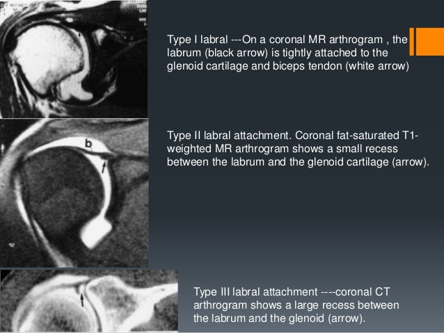
On mri the glenohumeral ligaments are best assessed in the presence of capsular distension which is produced if there is a large amount of joint fluid or contrast in the shoulder joint 22 37 43 44.

Bts love yourself tear concept photos. To rule out a labral tear an mri arthrogram needs to be ordered not an mri with contrast. The ighl can be confused with an anterior labral tear or a sublabral foramen where there is an anomalously high origin of its. To compare preoperative non contrast magnetic resonance imaging mri with arthroscopy findings in diagnosing labral and rotator cuff tears.
Clinical presentation patients with labral tears may present with a wide range of symptoms depends on the injury type which are often non. Magnetic resonance imaging of the shoulder. Rotator cuff tears the aber view is also very useful for both partial and full thickness tears of the rotator cuff.
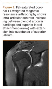
In addition there is faint extension of contrast material arrowhead into the superior labrum. Slap tears involve the superior glenoid labrum where the long head of biceps tendon inserts. An mri scan with contrast can take anywhere from 30 minutes to 90 minutes depending on the area of the body being scanned the agent used and the route of administration of the gbca.
Mris using oral gbcas may take up to two and a half hours requiring you to drink multiple doses and wait until the agent passes into the intestine. On images of the shoulder with the arm in a neutral position the torn labrum may be held in its normal anatomic position by the intact scapular periosteum which thereby prevents contrast media from entering the tear. Mri with or without intraarticular contrast administration is the preferred method for evaluating internal derangement of the shoulder.
A review of potential sources of. They can extend into the tendon involve the glenohumeral ligaments or extend into other quadrants of the labrum. When an mri with contrast is ordered contrast is injected into the vein while the arthrogram injects contrast directly into the joint under fluoroscopy guidance.
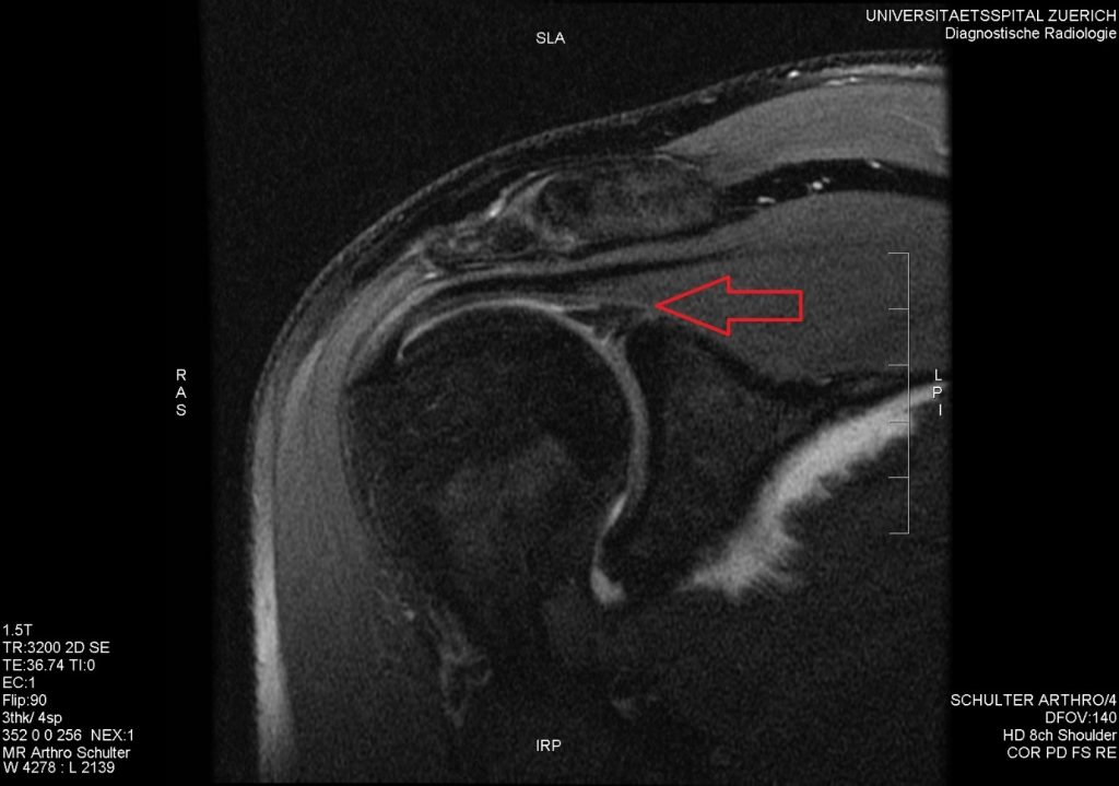
Slices were made in a transverse parasagittal and paracoronar orientation. A coronal mr arthrogram shows contrast material in the region of the glenoid labrum interface but the space between the glenoid and the labrum does not precisely follow the glenoid contour and is too wide to be considered normal. This means that mr arthrography with the arm in the neutral position may fail to detect the labral tear.
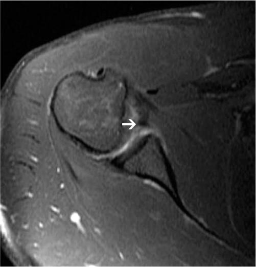
In the aber position the inferior glenohumeral ligament is stretched resulting in tension on the anteroinferior labrum allowing intra articular contrast to get between the labral tear and the glenoid. In the acute setting they are most frequently seen in. Glenoid labral tears are the injuries of the glenoid labrum and a possible cause of the shoulder pain.
More From Bts Love Yourself Tear Concept Photos
- Young Forever Bts Lyrics Romanized
- Jimin 2020 Filter
- Bts Love Yourself Her Concept Photos O Version
- Min Yoongi Y Su Hermano
- Bts Wallpaper Desktop Purple
Incoming Search Terms:
- Labral Tear Clinical Mri Bts Wallpaper Desktop Purple,
- Case Studies Portland Mri Siker Medical Imaging Bts Wallpaper Desktop Purple,
- Glenohumeral Instability Radsource Bts Wallpaper Desktop Purple,
- Acetabular Labral Tear Radsource Bts Wallpaper Desktop Purple,
- Shoulder Instability The Role Of Mr Arthrography In Diagnosing Anteroinferior Labroligamentous Lesions Our Experience At King Hussein Medical Center Bts Wallpaper Desktop Purple,
- The Radiology Assistant Shoulder Anatomy Mri Bts Wallpaper Desktop Purple,


/GettyImages-548557033-59ff685113f12900377707db.jpg)


