Torn Shoulder Labrum Mri Image, Shoulder Labral Tear Orthopaedic Neurosurgery Specialists
Torn shoulder labrum mri image Indeed lately has been hunted by consumers around us, perhaps one of you personally. Individuals now are accustomed to using the net in gadgets to see image and video information for inspiration, and according to the title of the post I will discuss about Torn Shoulder Labrum Mri Image.
- Shoulder Pain Clicking 3t Mri Shoulder Portland Mri Siker Medical Imaging
- The Radiology Assistant Shoulder Anatomy Mri
- Mr Arthrography Helps Labral Tear Patients Avoid Surgery
- The Radiology Assistant Shoulder Instability Mri
- Hill Sachs Deformity And Labral Tear Radiology Case Radiopaedia Org
- Arthrography Demonstrating Shoulder Labrum 3t Mri Shoulder Portland Mri Siker Medical Imaging
Find, Read, And Discover Torn Shoulder Labrum Mri Image, Such Us:
- Mri Arthrogram Glenoid Labral Tear Medical Imaging Mri Radiology
- Posterior Labrum Tear 3t Mri Arthrogram Shoulder Portland Mri Siker Medical Imaging
- Shoulder Labral Tear What You Should Know And What Surgeons Won T Say
- Superior Labral Anterior Posterior Lesions Of The Shoulder Current Diagnostic And Therapeutic Standards
- Slap Tears Orthoinfo Aaos
If you are searching for Jimin And V Friendship Necklace you've reached the perfect location. We have 104 images about jimin and v friendship necklace including images, photos, pictures, backgrounds, and much more. In these web page, we also provide variety of images out there. Such as png, jpg, animated gifs, pic art, logo, blackandwhite, translucent, etc.
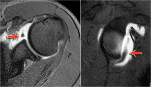
Mr imaging diagnostic criteria for labral tear versus anatomic variant.

Jimin and v friendship necklace. However there are several mr imaging characteristics that can aid. The process of diagnosing a labral tear is complicated by the various pitfalls in imaging diagnosis often present in the superior labrum 1734 which can make this a challenging task for the radiologist. One of the most frequent shoulder injuries is a rotator cuff tear.
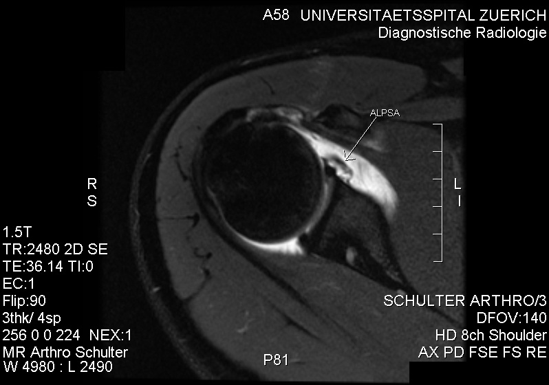
Unlike bankart lesions and alpsa lesions they are uncommonly 20 associated with shoulder instability 5. They can extend into the tendon involve the glenohumeral ligaments or extend into other quadrants of the labrum. It is a major cause of shoulder pain and weakness and accounts for a large amount of missed work sports and school due to injury.
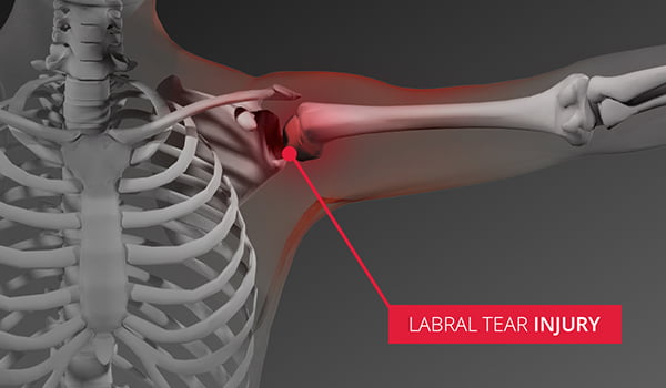
On images of the shoulder with the arm in a neutral position the torn labrum may be held in its normal anatomic position by the intact scapular periosteum which thereby prevents contrast media from entering the tear. To date no study has demonstrated that labrum tears lead to arthritis of the shoulder so even if. Glenoid labral tears are the injuries of the glenoid labrum and a possible cause of the shoulder pain.
Radiologist will often say possible labrum tear or labrum tear cannot be ruled out a suspected labrum tear is a very common finding on shoulder mri and again the finding has to be understood based upon your symptoms and your history. Clinical presentation patients with labral tears may present with a wide range of symptoms depends on the injury type which are often non. When an mri with contrast is ordered contrast is injected into the vein while the arthrogram injects contrast directly into the joint under fluoroscopy guidance.
To rule out a labral tear an mri arthrogram needs to be ordered not an mri with contrast. Over 2 million people in the united states suffer rotator cuff issues every year and need to have a rotator cuff mri. Mr arthrography is employed for the detection of subtle rotator cuff tears or labral pathology in patients with a negative conventional mri the assessment of the postoperative shoulder and the demonstration of communication between the joint and extra articular pathology such as a paralabral cyst.
In the aber position the inferior glenohumeral ligament is stretched resulting in tension on the anteroinferior labrum allowing intra articular contrast to get between. Labral tears the abduction external rotation aber view is excellent for assessing the anteroinferior labrum at the 3 6 oclock position where most labral tears are located. 2 direct mr arthrography distends the joint.
In the acute setting they are most frequently seen in.
More From Jimin And V Friendship Necklace
- Bts Persona Version 2 Concept Photos
- Bts Jimin Bt21 Chimmy
- Jimin Real Height
- If You Jungkook Caratula
- Meme Faces Bts Memes Love
Incoming Search Terms:
- Slap Tears And Biceps Tendon Injuries Huang Orthopaedics Meme Faces Bts Memes Love,
- Hip Labral Tears And Femoroacetabular Impingement A Frequent Cause Of Non Arthritic Hip Pain Derek Ochiai Md Nirschl Orthopaedic Center Meme Faces Bts Memes Love,
- Https Encrypted Tbn0 Gstatic Com Images Q Tbn 3aand9gcs72aqbjs4kxvkxcg9wa54wkcwyayh5yhishg Usqp Cau Meme Faces Bts Memes Love,
- Arthroscopic Treatment Of Posterior Shoulder Instability Orthopaedicsone Articles Orthopaedicsone Meme Faces Bts Memes Love,
- Glenohumeral Instability Radsource Meme Faces Bts Memes Love,
- Shoulder Mri Report Meme Faces Bts Memes Love,


